Research summary: shape analysis of Perthes’ disease deformity
This post is a summary of recent work in collaboration with Prof Andrew Anderson and Dr Joseph Mozingo at the University of Utah, and Dr Harry Kim at the Texas Scottish Rite Hospital for Children. It was recently published in the International Journal of Computer-Assisted Radiology and Surgery as:
Useful downloads:
The evaluation model from our study can be viewed by opening ‘EXTEND.xlsx’ using ShapeWorks. Please note that the surface files have been scaled up 3x, as this helped with particle distribution in ShapeWorks v6.3.0

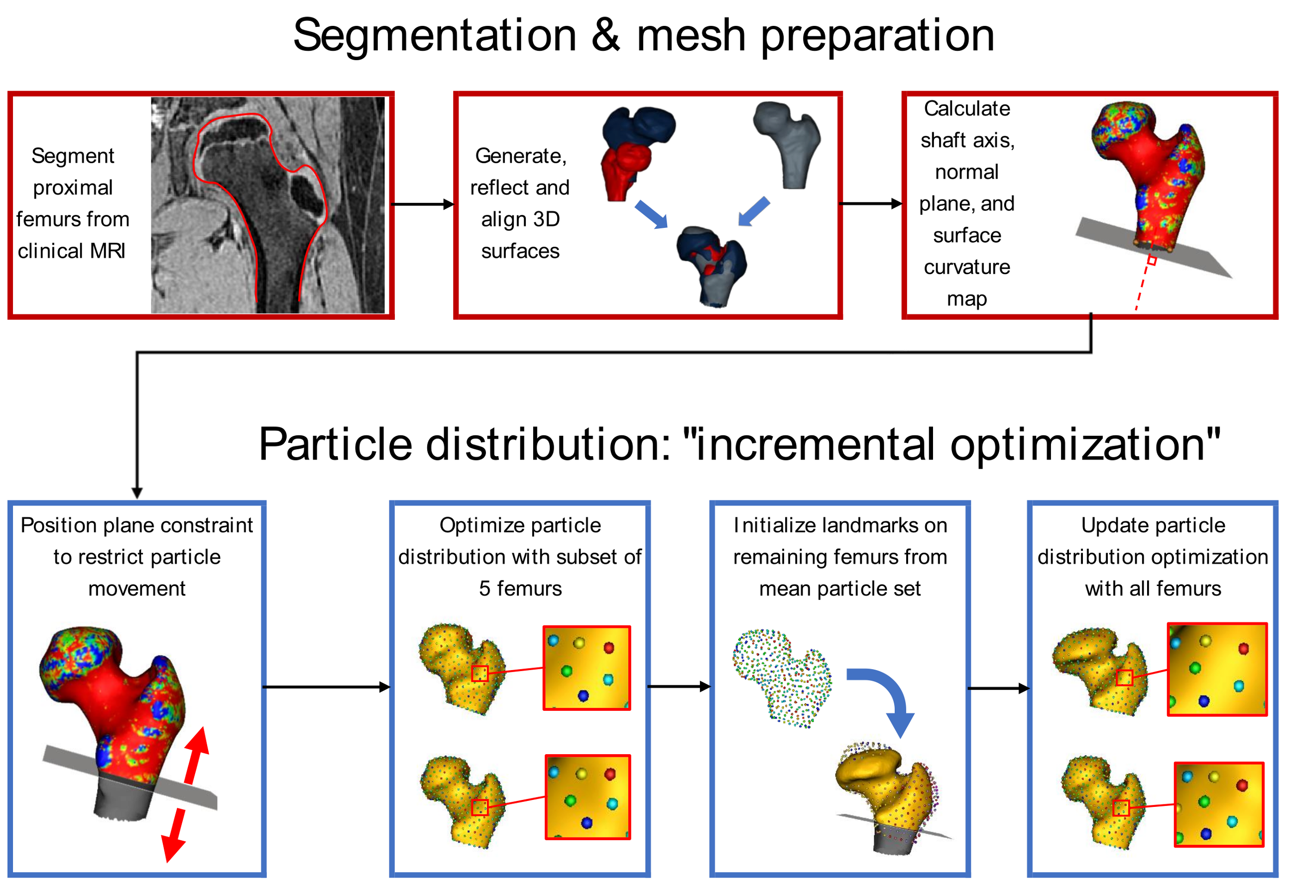
Our framework provides a step-by-step guide for making statistical shape models of Legg–Calvé–Perthes disease deformity.
Public summary
Perthes’ disease is a rare hip condition in children that can permanently change the shape of the hip joint. These shape changes are linked to problems like pain and arthritis later in life, and normally people with less rounded hips have a higher chance of developing these problems. Doctors and scientists measure these shape changes on X-rays to study how well different treatments for Perthes’ have worked.
However, measuring hip joint shape from X-rays is not ideal. The main downside is that they are 2D pictures, and might not be able to show some of the differences between complicated 3D hip deformities. We can take 3D pictures of the hip using MRI or CT, but at the moment we don’t have a way to measure the overall 3D shape of the hip joint in Perthes’ disease.
In this study I collaborated with researchers from the University of Utah and the Texas Scottish Rite Hospital for Children to develop a framework (basically a step-by-step guide) for making statistical shape models (SSMs) of hips with Perthes’ deformity. These are mathematical methods that take in a group of shapes and output a small number of variables that describe the main ways those shapes vary across the group. For example, if you produced an SSM from randomly picked coffee mugs, the first mode might represent the size of the mug from espresso to venti, the second might correspond to the roundness of the mug’s base, and so on.
Making an SSM of 3D Perthes’ deformity is difficult because a) hips with Perthes’ deformity vary a lot in shape and b) Perthes’ is rare and 3D pictures are not often taken, so it’s hard to collect a large enough dataset to produce an accurate model. For our framework, we used a new way of producing SSM that makes it better at dealing with a), and we tested the framework on a small group of MRI scans from an older study. It performed really well, and accurately represented shape changes that are common in Perthes’!

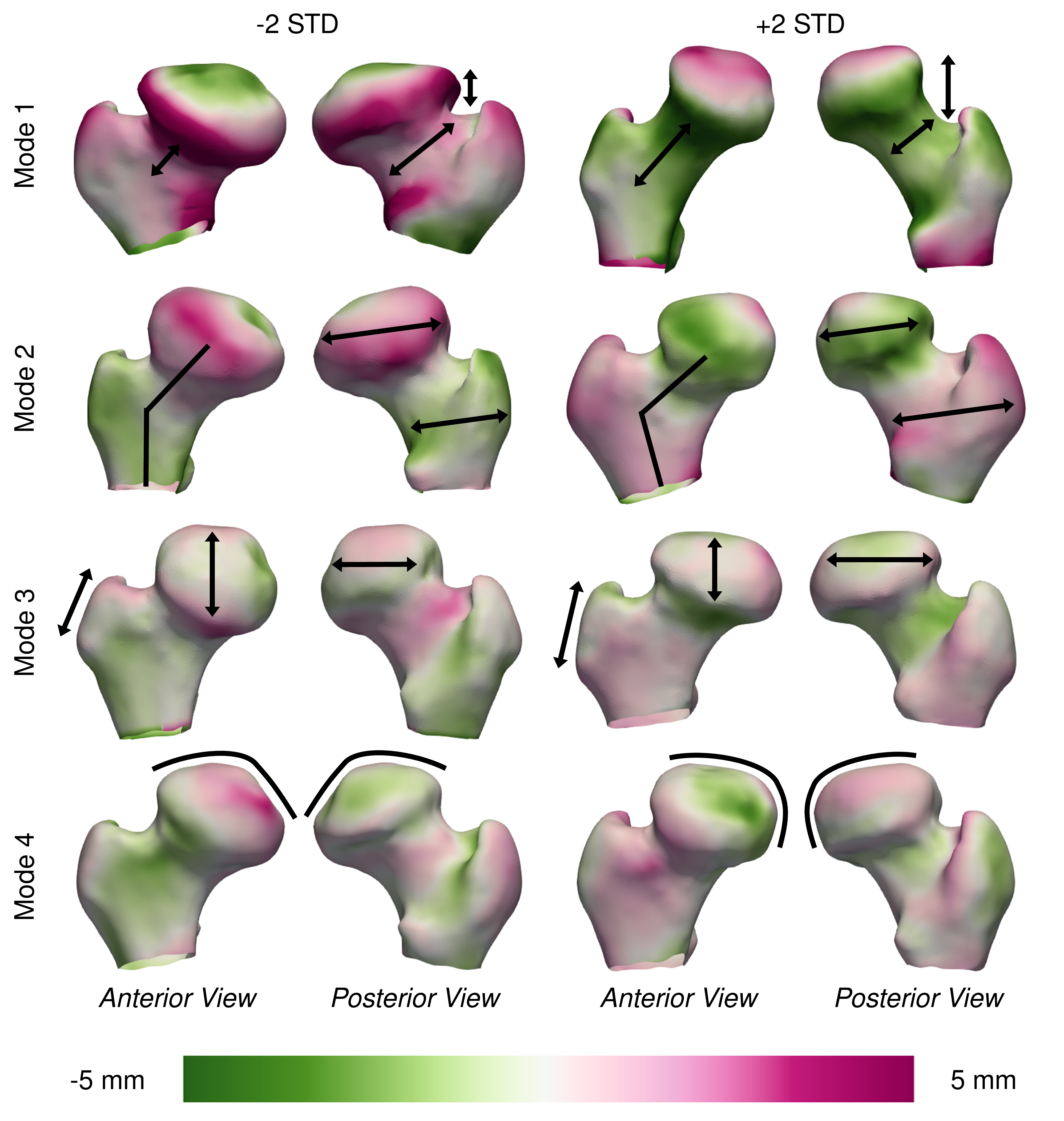
We can see shape changes captured by our evaluation model by varying the four most important modes. The strength of the green and magenta colours represent how far the shape’s surface has moved from the average.
The framework also makes it easy to add more data to an existing SSM, which we hope will encourage doctors and scientists studying Perthes’ deformity to add to our current dataset over time, making it better and better at describing all kinds of Perthes’ hips. There’s still a lot to do, but eventually this could become a very useful tool for researchers, providing an accurate representation of overall deformity in 3D that could then be compared with clinical measurements like X-rays and outcomes for patients like pain or quality of life.