It’s been just over a year now since I posted about my transcontinental Odyssey from Vancouver to Boston, where I casually mentioned that I was a postdoctoral research fellow at Northeastern University “messing around with hips for a couple of years”. As this position has drawn to a premature close (more on that later), I thought I might expand on that aside and do a bit of a retrospective of the last year and a bit.
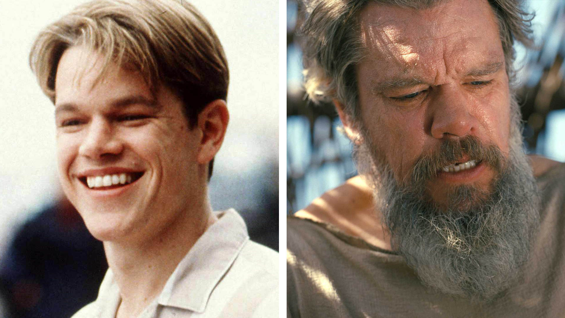
Me before (left) and after (right) driving across Canada.
Disclaimer: some of the work I cover is ongoing so I’ll be a little sparse on the details. Rest assured that anything that gets published will get a summary on this site as always!
Why Boston?
My then-fiancée (now wife) and I both finished up our PhDs at UBC at about the same time, and thanks to the specifics of her anticipated career path, she got priority on choice of postdoctoral research labs and ended up choosing Harvard. Thankfully, being a biomedical engineer in Boston is like looking for snacks at Market Basket: you won’t lack for options.
This is how, after a thankfully very brief job hunt, I ended up at Northeastern University’s Institute for Mechanobiology. In early July 2025 I started my postdoc with Prof. Sandra Shefelbine as my primary research supervisor, although the position itself was funded for two years by the Institute (foreshadowing). This unique setup gave me a whole lot of autonomy with my research focus, which I planned to take advantage of.
Autumn Fall
After spending most of the summer in IT and onboarding purgatory (thankfully there were some grant applications I could help out with to keep me busy), things really got going in the fall. I put a lot of effort into making myself useful in the lab and the Institute, and I was glad to see that I could share and adapt my PhD work to help out Sandra’s grads.
This involved some stuff I was very at home with—hips! I helped a new PhD student, Laura Wang, get set up with the Healthy Hips project, a collaboration Sandra had with Prof Cara Lewis at Boston University. Her specific focus was to detect early changes in cartilage health in female athletes using T1ρ imaging, allowing me to generously donate and troubleshoot my existing analysis code.1 I also dabbled a little in comparing hip joint centre calculations between MRI scans and motion capture data; sadly the grant application that could have turned this into a full project was unsuccessful.2
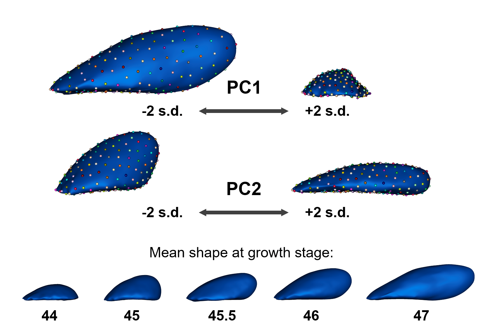
Helping out with PhD student Soha Ben Tahar’s work on axolotl limb development was a little further outside my comfort zone. Her work investigates how the distribution of tissue mechanical properties and biochemical signals causes the highly-retained structure of vertebrate limbs: one bone close to the body, two bones further down, and then many many bones at the very end. Part of this work involves 3D fluorescence imaging of key markers during the very earliest stages of limb growth, when the limb starts “budding” out from the torso, and in order to compare the distribution of these markers between individual buds they need to be mapped onto a common morphological reference for that growth stage.
What can efficiently describe highly variable shape populations and provides a common surface particle representation to enable morphing between examples? That’s right, statistical shape models! The limb buds were an even bigger challenge than Perthes’ hip deformity—as can be seen with the huge shape change described by the first principal components—but with some particle-initialization hacks I managed to construct a model that stayed smooth through all the growth stages of interest. The model could then be used to automatically place landmarks on new limb buds for reference morphing.
As an Institute for Mechanobiology postdoc I took my Institute responsibility very seriously as well, taking the time to familiarise myself with the website and maintain the events page, as well as setting up a Bluesky and LinkedIn profile and trying out a “skills workshop” event — something that the Institute could provide for trainees that wasn’t just another seminar series.
Between all these activities I somehow found time to do my own research. My main focus was something that has been frustrating me for years: segmentation! A word to put a shiver down any co-op, research assistant, or grad student’s spine. Segmentation is the process of splitting 2D or 3D medical images into regions (or “segments” — see?) corresponding to different tissues or anatomical structures, and is critical for a wide variety of biomedical analyses. For example, in Laura’s T1ρ studies she needs to segment the thin cartilage in the hip joint in order to run her analysis on only that specific tissue, and in my earlier biomechanical simulation of the hip with Perthes’ deformity, the patient-specific model of hip anatomy was created by segmenting an anatomical MRI scan.
Segmentation is also extremely time consuming. In the same Perthes’ simulation study, it took me about 20 hours (if I recall correctly) to segment the single pair of hips I included. Many very clever people have come up with many very clever ways to automate this process, most prominently including machine-learning approaches such as nnU-Net. These work great for relatively uniform image types like CT, but the huge variety of MRI scans and the lack of large musculoskeletal MRI datasets mean bespoke segmentation models often need to be trained for each study; a model trained on one kind of MRI scan often doesn’t work on another. So how do you get a training dataset? More manual segmentation, of course!
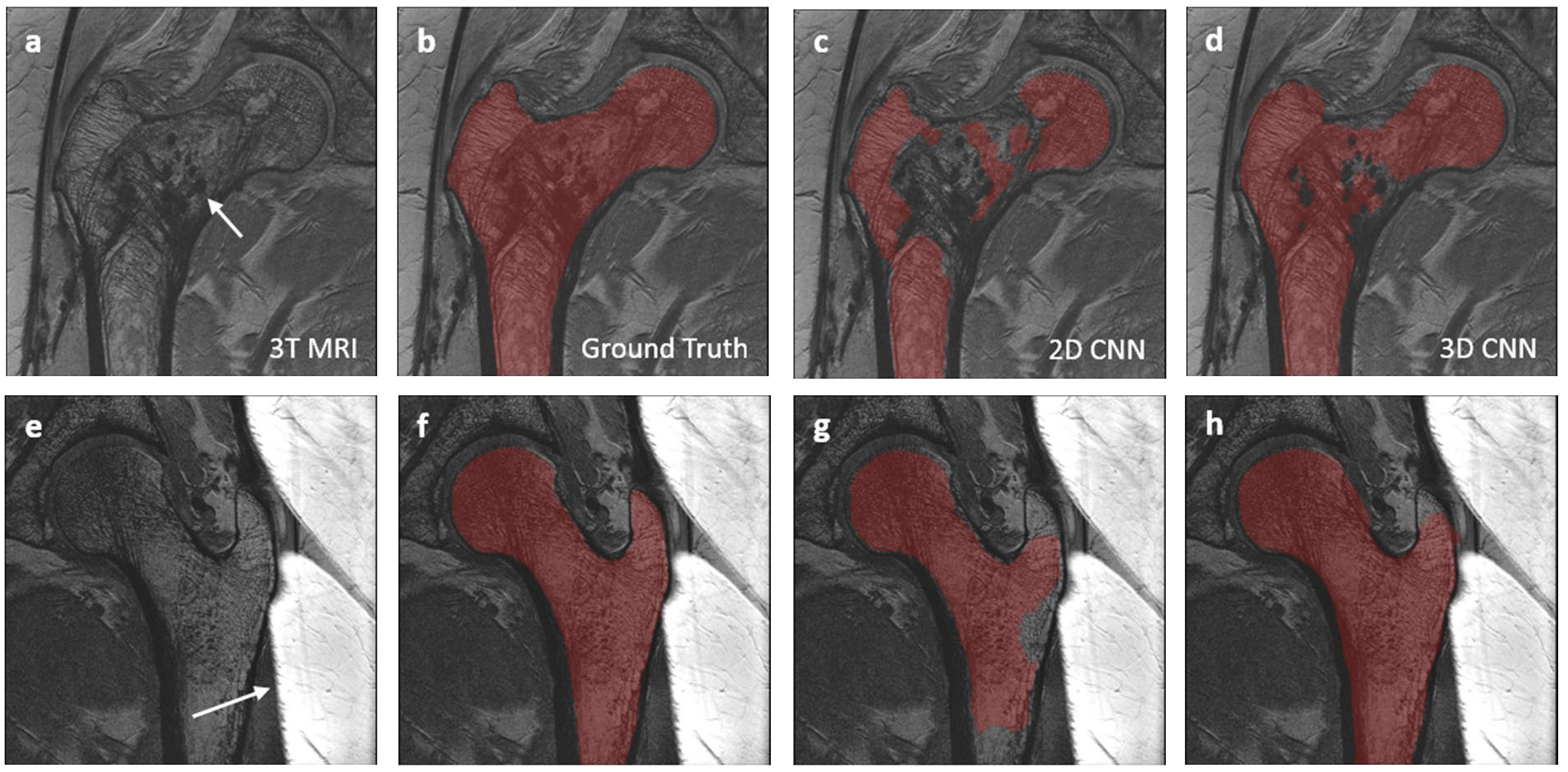
I wanted to see whether I could once again leverage my PhD experience to improve the transfer of automatic segmentation models between different kinds of image by bringing in statistical shape models as an initial step. The idea was that a rough segmentation estimate in the correct location and with the right kind of shape would help to prevent the kinds of “anatomically infeasible” failures that happen when a machine-learning segmentation model encounters unexpected data. The specific details I’ll leave for another time, but as of writing the approach shows a lot of promise.
Examples of “anatomically infeasible” segmentation failures upon encountering anatomical abnormalities (bone cysts, top row) and image artifacts (wraparound, bottom row) in MRI scans. From Deniz et al. 2018, Creative Commons BY 4.0.
Needless to say I kept myself busy. By the end of the fall I was starting to feel like I was really settling in and building momentum, I was feeling ready for 2026 to be a bumper year. But then…
Winter
Only in the first week back after the holidays, I found out that the University was pulling all funding for research institutes. You know, the funding that paid my salary? My two-year position was now going to be a one-year position unless we could scrape something together fast.
As you can imagine, this hit my motivation quite badly at the expense, mainly, of my self-assigned Institute responsibilities. So almost as soon as the socials were set up they went quiet, I stopped proactively asking around for news updates, and the first skills workshop was also the last. This last one was a big shame, because we had a lot of really good feedback and ideas about how to move forwards. Instead, I put all of my effort into two things: my hybrid shape/machine-learning segmentation idea (now called ADAPT-seg: Agnostic, Deep-learning And Prior-guided Tissue segmentation), and applying for GRANTS GRANTS GRANTS based on the ADAPT-seg work.
A musical interlude
This might seem extracurricular but I’m counting it as work-related: I joined a choir! The Longwood Chorus is made up of medical students and professionals as well as allied researchers so it’s a very sciencey bunch.
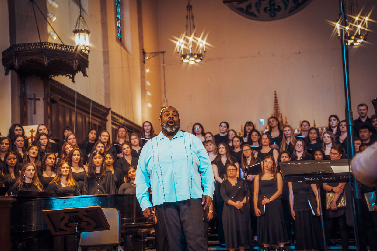
Me (back row, far right) entranced by Grammy-nominated countertenor Reginald Mobley during Longwood Chorus’ performance of Chichester Psalms, May 2026. Photo credit: Ziggsphoto (gallery)
They do two main concerts a year with a pretty broad and challenging repertoire, as well as a scattering of other run-outs with smaller groups. Having performed at the American Academy of Arts and Sciences induction ceremonies in both 2025 and 2026, I can now say that I’ve sung for Anderson Cooper, José Andrés and Jodie Foster!
Summer
Spring in Boston is basically two weeks long. It felt like we went from snow on the streets to shorts and sunbathing within the month of April.
The limbo feeling intensified as my nominal Jun 30th end date drew closer and closer, but with only two weeks to go I had a stay of execution: one of our short-term grant applications had been successful! This was a pilot grant from the Multicenter Osteoarthritis Study (MOST), which covered my salary until October. Other more long-term applications are still pending, but would allow me to come back next year if successful.
Why oh why, IWOAI?
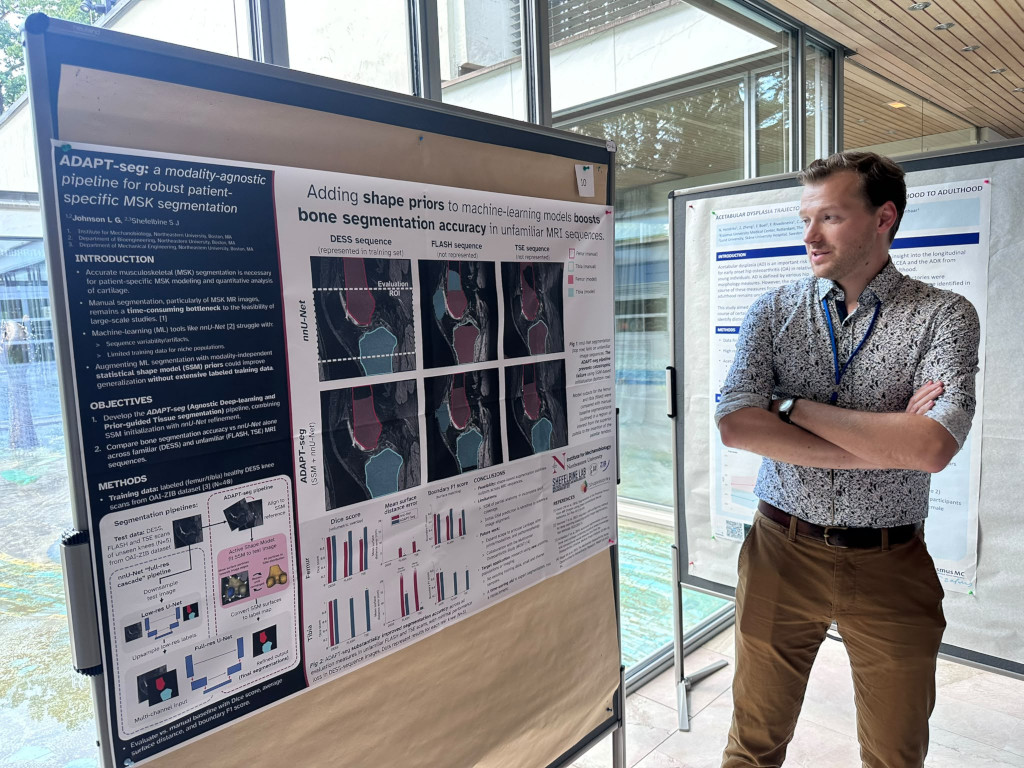
The MOST pilot grant was doubly relieving, as it meant I could attend the International Workshop on Osteoarthritis Imaging (IWOAI—pronounced eye-why) in a sweltering Munich at the end of June,3 without the looming threat of unemployment on my return. This is a great little conference with a strong networking and social factor, and I was able to present the latest ADAPT-seg progress with a poster and link up with John Lynch from MOST to chat strategy.
I look very sceptical of my own results, don’t I.
For more of my IWOAI experience and thoughts, check out the big old thread I made on Mastodon and Bluesky.
From my return until the MOST money ran out a week ago today—with a short break to get married—the focus was entirely on getting ADAPT-seg into a suitable shape. This involved adding cartilage where previously we’d only attempted to segment bone, automating the remaining manual steps, and packaging everything up in a somewhat rational way for future users. I’ve also got the ball rolling on writing up as well, but am waiting on grant news before making any firm decisions about scope. In the mean time, the ADAPT-seg code is on Github already, you know, just in case.
And that’s about it! I really enjoyed my time at Northeastern and was disappointed to have it cut short. For now, though, I’m making the most of my free time (surely not by writing this blog? - L) and crossing my fingers that I can pick up where I left off next year.
Itself stolen/adapted from Carly Jones, my PhD predecessor at UBC. ↩︎
The application in question was a second attempt: it had been unsuccessful the year before, was tidied up and resubmitted as a much more solid project. This time around, it scored even lower! Dare I suggest that the shifting priorities of the US government might have tipped the scales against women’s health research…? ↩︎
I must also admit responsibility for Germany’s terrible performance at the World Cup. Normally my sporting curse only brings down teams when I watch their games (see England in the semi), but this time it appeared that just being physically present in the country was enough. ↩︎
Comments
You can use your Mastodon or other Fediverse account to comment on this post by replying to this thread.

Comments
You can use your Mastodon or other Fediverse account to comment on this post by replying to this thread.
Alternatively, you can reply to this Bluesky thread using a bridged Bluesky account.
Learn how this is implemented here.